Petri Dish Mold Identification: Agar Colony Photos & Chart
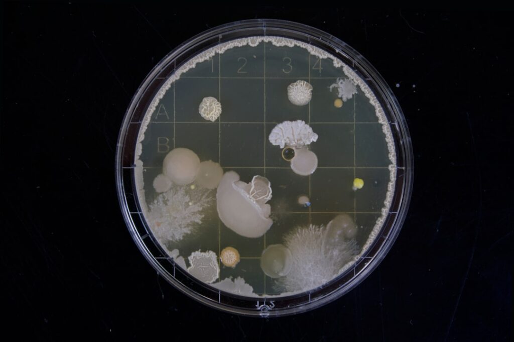
Home mold kits make a simple promise: leave a dish open, let colonies grow for a few days, then compare them to petri dish mold identification photos online and name what is growing in your house. Colony photos can be genuinely informative about what kind of fungus you might be looking at. They cannot tell you which species it is, how much mold is in your home, or whether anyone is being exposed. This guide gives you a descriptive identification chart for the eight genera most often asked about, explains what black mold usually looks like on a plate (and why it often does not show up at all), and covers how to photograph and dispose of plates safely. It summarizes EPA and CDC guidance current as of 2026. It is not medical advice; talk to a physician about symptoms and a qualified professional about testing decisions.
Part of our Types of Mold guide — start there for the full picture →
Is this mold, asbestos, or water damage? Get a free instant screen
Upload a clear photo of the suspect area. You'll get an instant AI screening opinion and what to do next. This is screening guidance, not a professional determination.
Screening guidance only. AI can be wrong. Confirm asbestos, mold type, or lead with lab testing or a licensed professional before acting.
Can You Identify Mold From a Petri Dish Photo?
Partly. A colony’s color, texture, growth speed and the color of its underside (the “reverse”) can suggest a likely genus, in the same way a leaf can suggest a tree family. Experienced lab staff use those features as a first clue. They do not stop there, because many molds look alike as colonies and the same mold can look different from plate to plate.
What changes a colony’s appearance:
- The growth medium. Different agars produce different colors and textures from the same organism.
- Temperature and light. A plate on a warm kitchen counter grows differently from one in a cool basement.
- Age. Many molds start white and only develop their characteristic color as they produce spores. A three-day photo and a seven-day photo of the same colony can look like two different molds.
- Crowding. Fast growers overrun slow ones, and colonies touching each other can change shape and color.
Most online galleries do not say which medium, temperature or incubation time produced the photo. Matching a homemade plate to a random image is closer to a hint than an identification.
How a Settle Plate Kit Works
Most home kits use the settle plate method. The dish contains a nutrient gel, often a potato dextrose or similar fungal agar, sometimes with an additive that slows bacteria. You remove the lid for a set time, usually somewhere between several minutes and an hour depending on the kit, then close it and wait. Any viable spores that dropped onto the gel during that window can germinate and grow into visible colonies over the next two to seven days.
Three details shape every photo you will take later:
- Only viable, culturable spores grow. Dead spores and fragments, which can still matter for allergies, leave no colony. Some molds also grow poorly on general media.
- Heavy spores settle; light ones may not. Gravity decides much of what lands, so the plate is a biased sample of the air, not a measurement of it.
- Whatever grows fastest wins. A single Mucor or Trichoderma spore can cover a plate before slower molds get started.
Getting a plate that is worth photographing
If a kit is going to be used, a few habits make the result less random. Follow the kit’s exposure time exactly. Place the dish at about table height in the middle of the room, away from fans, open windows and pets. Run a second plate outdoors at the same time, under cover, as a rough comparison; the EPA’s mold course notes that after remediation, the types and concentrations of mold indoors should be similar to those in the local outdoor air, and that omitting outdoor control samples is one of the common sampling problems. Keep both plates at the same room temperature while they incubate, and photograph both on the same days. An indoor plate that looks very different from the outdoor one, especially with a genus that is scarce outside, is a reason to look harder for moisture. A plate that looks like the outdoor one is weak evidence of anything.
Petri Dish Mold Identification Chart
The chart below describes how colonies of common indoor and outdoor genera typically appear on general-purpose fungal media at room temperature. These are textbook tendencies, not rules. Every row has exceptions, and several genera can look nearly identical at certain stages.
| Genus | Typical colony front (color and texture) | Typical reverse (underside) | Growth speed on a plate | Common indoor sources |
|---|---|---|---|---|
| Aspergillus | Powdery to velvety. Color depends on species: blue-green, yellow-green, yellow, brown or jet black, often with a white edge while young. | Usually pale, cream or yellowish; some species darker | Moderate to fast; visible in a few days | House dust, HVAC systems, damp building materials, stored food |
| Penicillium | Velvety to powdery blue-green or gray-green, often with a white margin and radial grooves | Pale, cream or yellow; varies by species | Moderate; often one of the first to cover a plate | Water-damaged drywall, wallpaper, carpet, dust, spoiled food |
| Cladosporium | Compact, velvety, olive-green to dark brown or nearly black, sometimes with a slightly raised center | Dark olive to black | Slow to moderate | Outdoor air drawn indoors, window frames and sills with condensation, bathroom surfaces, HVAC |
| Stachybotrys | Starts white, becomes dark gray to black and sooty or slightly slimy as it sporulates | Pale to dark, sometimes with a darker pigment | Slow; often outcompeted on settle plates and may not appear at all | Chronically wet cellulose: the paper face of drywall, ceiling tiles, cardboard |
| Alternaria | Woolly to cottony, gray-green to dark olive or black, sometimes with a lighter edge | Dark brown to black | Fast | Outdoor air, window frames, showers, damp textiles |
| Mucor / Rhizopus | Tall, fluffy “cotton candy” growth, white to gray; Rhizopus develops visible black dots (spore sacs) | Pale or white | Very fast; can fill a plate and lift the lid in a few days | Food, soil, house plants, damp organic debris |
| Fusarium | Cottony, white at first, often turning pink, salmon, lavender or purple | Often pink, red, purple or pale | Fast | Water-damaged materials, humidifiers, soil and plants |
| Trichoderma | White and cottony at first, then bright to dark green patches, often in rings or tufts | Pale to yellowish | Very fast | Wet wood, damp drywall and wallpaper, soil |
The CDC lists Cladosporium, Penicillium and Aspergillus as the most common indoor molds, so those are the colonies most homeowners see first on an indoor plate. For deeper profiles of each, see our guides to Aspergillus, Penicillium mold, Cladosporium and Stachybotrys. A printable visual version lives in our petri dish mold identification chart.
Reading the chart honestly
Use the chart to narrow possibilities, not to settle them. A blue-green velvety colony could be Penicillium or one of several green Aspergillus species. A dark olive colony could be Cladosporium, Alternaria, or another dark fungus not listed. Fusarium and some unrelated fungi can share pink tones. Without looking at spores and spore-bearing structures under a microscope, these cannot be reliably separated, which is why the chart includes no “this is definitely” column.
What Does Black Mold Look Like in a Petri Dish?
“Black mold” is the most searched phrase in this topic, and also the most misunderstood. The EPA’s mold course is direct: “black mold” is not a species or a specific kind of mold, and neither is “toxic mold.” The media sometimes use those terms for molds that may produce mycotoxins, or specifically for Stachybotrys chartarum.
On a settle plate, black or near-black colonies are very common, and most are not Stachybotrys. Dark colonies on a home plate are more often:
- Cladosporium, which is olive to black, compact and velvety, with a black reverse.
- Alternaria, which is woolly and gray-black to olive-black.
- Aspergillus niger and related species, which form powdery, jet-black spore heads on a lighter, often yellowish base, a “pepper on cream” look.
- Rhizopus, which looks white and fluffy but becomes dotted with black spore sacs.
Stachybotrys itself grows slowly, needs wet cellulose, and does not release spores into the air easily, so it often does not land on an open plate at all, and when it does, faster molds tend to overrun it. A home plate with no black colonies does not rule out Stachybotrys in a wall, and a plate full of black colonies does not mean it is present. Our pages on black mold in a petri dish, black mold petri dish pictures and what black mold looks like in a petri dish show side-by-side examples.
Different Types of Mold in One Petri Dish
A home plate almost always grows several kinds of mold at once, often alongside bacteria and yeasts. That mix is normal. The EPA notes that mold spores are found both indoors and outdoors and that the number in the air fluctuates from season to season, day to day and even hour to hour. An open dish samples whatever settles during that window.
Ways to tell colonies apart on a mixed plate:
- Mold versus bacteria and yeast. Mold colonies are fuzzy, powdery or cottony because they produce threadlike filaments. Bacteria and yeasts usually form smooth, shiny or creamy dots that look wet.
- Each distinct texture is a candidate. Count colonies by appearance: velvety green, cottony white, compact black, and so on.
- Watch over several days. Photograph at the same time daily. Color development and growth speed are part of the chart above, and a time series shows both.
- Look at the reverse. Flip the sealed dish and photograph the underside. Reverse color is one of the more useful clues for dark molds.
Our broader mold petri dish guide covers kit setup, and mold colors explains why color alone misleads.
How to Photograph Petri Dish Mold Safely
Photos are the safest way to study a plate, because they let you zoom in without exposure. The point is to look, not touch. Growing dense colonies concentrates spores, and opening the lid releases them into the room you live in. The CDC notes that people with asthma or who are allergic to mold may have severe reactions, so keep plates away from anyone in that group.
Keep the dish sealed
- Once the exposure period ends, close the lid and tape it shut around the edge with clear tape or paraffin film if the kit includes it.
- Never open the lid to sniff, touch or poke a colony, even with a cotton swab.
- Store plates lid side down, if the kit instructions agree, so condensation does not drip onto the colonies.
Getting a useful photo
- Use indirect daylight or a lamp at an angle to avoid glare off the lid.
- Shoot the front and the reverse, with a ruler or coin beside the plate for scale.
- Use the phone’s macro mode or a clip-on macro lens; avoid pressing the phone against the lid.
- Label each plate with date, room and exposure time, and keep that label visible in the photo.
Apps that promise to name the mold from your photo face the same limits as a gallery match; see our review of the mold identification app category.
Dispose of plates properly
Do not open the plate to clean and reuse it. Keep it taped shut, place it in a sealable plastic bag, seal a second bag around it, and put it in the household trash. Follow the kit’s own disposal instructions if they differ. Wash your hands after handling plates, even sealed ones, and wipe the surface where they sat.
What the Photos Can’t Show
Even a perfect photo of a perfectly grown plate leaves the important questions open.
- Species. Reliable identification needs microscopy of spores and spore-bearing structures, or a culture analyzed by an accredited laboratory, sometimes with molecular methods. The EPA’s mold course notes that bulk samples can be examined using microscopes in the lab; a phone camera cannot substitute for that.
- Amount. A settle plate does not measure how many spores are in the air. It shows what happened to land and grow during the exposure time.
- Source. A colony on the plate does not tell you where it came from: a wall cavity, a houseplant, or someone’s shoes.
- Exposure or health risk. No plate result tells you whether anyone is being exposed at a level that matters. Health questions belong with a physician.
- Hidden growth. Molds growing inside walls, under carpet or in ductwork may never reach the plate.
Our mold microscope guide explains what lab analysts actually look for, and mold identification walks through the professional process from sample to report.
How a Lab Identifies Mold
When identification actually matters, the work moves to a laboratory. The EPA advises that sample analysis should follow analytical methods recommended by the American Industrial Hygiene Association (AIHA), the American Conference of Governmental Industrial Hygienists (ACGIH), or other professional organizations. Labs doing environmental mold work commonly hold AIHA laboratory accreditation for that purpose, which is worth asking about before any sample is sent.
The main approaches are:
- Direct microscopy. A tape lift, swab or bulk piece of material is examined under a microscope. The analyst looks at spore shape, size, color and the structures that produce them. This is fast and identifies many molds to genus, but not usually to species.
- Spore trap analysis. A pump draws a measured volume of air across a sticky slide, and the analyst counts and classifies the spores. Because the air volume is known, results can be reported as spores per cubic meter and compared with an outdoor sample taken at the same time.
- Culture under controlled conditions. Samples are grown on specific media at set temperatures, then each colony is examined under the microscope. This can identify some molds to species and confirms they are viable, but it takes a week or more.
- Molecular methods. Some labs use DNA-based tests for targeted species. Results depend heavily on how the sample was collected and which species the test covers.
Each method answers a different question, and none of them replaces a site inspection that finds the moisture. A good report pairs lab numbers with an indoor-versus-outdoor comparison and a plain-language interpretation from the professional who designed the sampling.
Why Settle Plates Mislead and What to Do Instead
The EPA’s mold testing page states that, in most cases, if visible mold growth is present, sampling is unnecessary. It also notes that no EPA or other federal limits have been set for mold or mold spores, and that sampling should be conducted by professionals with specific experience designing sampling protocols and interpreting results. A home settle plate falls short on each of the points the EPA’s mold course lists as common sampling problems: too few samples, inconsistent protocols, contamination, and omitted outdoor control samples. The course adds that inadequate sample plans may generate misleading, confusing and useless results.
The CDC puts the practical point plainly: if you see or smell mold, you should remove it, and you do not need to know the type of mold. That is the most useful sentence for anyone holding a plate of colorful colonies.
A better order of operations
- Look and smell. Visible growth or a persistent musty odor matters more than a plate result.
- Find the moisture. The EPA’s guidance is that the key to mold control is moisture control, and that water-damaged areas should be dried within 24 to 48 hours. Check plumbing, windows, roof leaks, basements and crawl spaces.
- Control humidity. The EPA recommends keeping indoor humidity below 60%, ideally between 30% and 50%, and the CDC recommends no higher than 50%. Front Range homes are usually dry in winter, but basements, swamp coolers and bathrooms can still run humid.
- Clean small areas properly. The EPA indicates that areas under about 10 square feet can usually be handled by the homeowner following its guidelines; larger areas or heavy water damage call for experienced help.
- Test professionally when it would change a decision. Real estate transactions, unexplained odors, post-remediation clearance, or health concerns are reasons to bring in someone who will take paired indoor and outdoor samples and send them to an accredited lab. See our guide to air sampling for mold.
If a DIY kit is still on your list, our review of the at-home mold test kit options explains which kits include lab analysis and which are settle plates only, and the mold test kit shop lists current products. For background on the mold groups in this chart, see types of mold, our mold identification chart for growth on walls rather than plates, the mold types hub, and the full mold inspection and testing guide.
References
- Mold Testing or Sampling — U.S. Environmental Protection Agency
- A Brief Guide to Mold, Moisture and Your Home — U.S. Environmental Protection Agency
- Mold Course Chapter 1: Introduction to Molds — U.S. Environmental Protection Agency
- Mold Course Chapter 3: Finding Mold and Moisture — U.S. Environmental Protection Agency
- Mold — Centers for Disease Control and Prevention
Frequently asked questions
How do you identify mold in a petri dish?
Note each colony's color, texture, growth speed and the color of its underside, then compare those features to a reference chart. That narrows the possibilities to a likely genus at best. Confirming the genus or species requires microscopy or culture analysis by an accredited laboratory.
What does black mold look like in a petri dish?
Many dark colonies appear on home plates, and most are common molds such as Cladosporium, Alternaria or Aspergillus niger. Stachybotrys, often called black mold, grows slowly and frequently does not appear on settle plates at all. The EPA notes that black mold is not a species or specific kind of mold.
Is it safe to open a petri dish with mold growing on it?
No. Opening the lid releases concentrated spores into your home. Keep the plate taped shut, photograph it through the lid, then double-bag it and put it in the trash. People with asthma or mold allergies should avoid handling plates.
Do petri dish results show how much mold is in my house?
No. A settle plate only shows which viable spores happened to land and grow during the exposure time. It cannot measure airborne concentration, find hidden growth, or show whether anyone is being exposed. Professional sampling with outdoor comparison samples is needed for those questions.
Should I test mold if I can already see it?
Usually not. The EPA states that in most cases, if visible mold growth is present, sampling is unnecessary. The priority is to fix the moisture problem and clean or remove the growth safely.
Got a plate full of colonies and no clear answer? Denver-area and Front Range homeowners can ask us to connect them with a vetted local inspector who can find the moisture behind the mold.
DIY mold test kits
A test kit can confirm whether a suspect patch is mold and (with lab analysis) what type — though visible mold over ~10 sq ft generally warrants a professional.
| Product | Why | Buy |
|---|---|---|
 DIY Mold Test (lab included) | Surface + air-sampling options. | Amazon — $17.95 |
 Healthful Home 5-Minute Mold Test | Rapid screening strips. | Amazon — $35.47 |
 Mold Test Plates (DIY petri dishes) | Settle-plate style sampling you culture at home. | Amazon — $22.99 |