Diagnosis of Lead Poisoning: Physician Pathway and Home Source
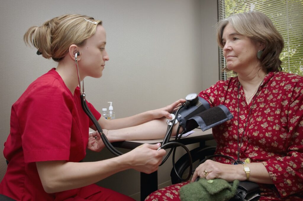
Diagnosis of lead poisoning is a medical decision made by a physician based on blood lead level (BLL) testing, clinical symptoms, exposure history, and in significant cases additional diagnostic workup including neurological and hematological assessment. No home test diagnoses lead poisoning in a person; home tests identify lead in paint, dust, water, or soil. The medical pathway and the home-source pathway run in parallel — both are usually needed to fully resolve a case. This guide summarizes CDC, NIH, and EPA guidance current as of 2026 and is intended as background for conversations with a physician. It is not medical advice. Anyone with suspected lead exposure should consult a licensed healthcare provider.
Part of our Lead Paint & Lead Testing guide — start there for the full picture →
Asbestos or biohazard concern? Speak with a certified contractor.
📞 Call (877) 742-8496Diagnosis of Lead Poisoning: The Clinical Framework
A physician evaluating a possible lead poisoning case typically follows a standardized clinical sequence. Step one is taking an exposure history — occupational risks, home age (pre-1978 housing is the key threshold), known renovation work, hobby exposures, drinking-water source, and dietary or cultural risk factors. Step two is symptom evaluation. Step three is venous blood lead level testing with confirmation if the first sample is elevated. Step four, for elevated BLLs, is additional workup that may include hemoglobin and hematocrit, iron studies, zinc protoporphyrin (ZPP) or erythrocyte protoporphyrin (EP), renal function, and neurological assessment.
The diagnostic threshold for “lead poisoning” varies by patient age and clinical context. The CDC currently uses a blood lead reference value of 3.5 μg/dL for children under six and tracks adult elevated levels through NIOSH surveillance. A BLL above this reference value combined with a plausible exposure history confirms a diagnosis of elevated blood lead level. Whether a particular case is also labeled “lead poisoning” depends on the level reached and the presence of clinical symptoms. The CDC childhood lead poisoning prevention program details the screening and follow-up algorithm.
Blood Lead Level Testing
The blood lead level test is the cornerstone of diagnosis. A venous blood sample is drawn into a lead-free royal-blue-top tube and analyzed by a clinical laboratory using ICP-MS, atomic absorption, or anodic stripping voltammetry. Results report in micrograms per deciliter and are usually available within several business days. Initial screening for children may use a capillary fingerstick draw, but any elevated capillary result is confirmed with a venous draw before treatment decisions are made.
BLL is a snapshot of recent exposure plus released bone stores, not a cumulative dose measurement. A patient with chronic past exposure may show a moderate current BLL while carrying significant bone-stored lead. Repeat testing at 1- to 3-month intervals tracks the trajectory and confirms whether exposure has stopped. The standard reference framework treats <3.5 μg/dL as low risk for general adult population, 3.5-10 μg/dL as elevated requiring follow-up, 10-25 μg/dL as significant, 25-45 μg/dL as concerning, 45-80 μg/dL as triggering possible chelation, and >80 μg/dL as medical emergency.
Neurological Assessment in Children
Pediatric lead poisoning produces neurological effects that may persist even after exposure stops. A physician evaluating a child with elevated BLL typically refers for developmental screening, cognitive assessment, and in some cases neuropsychological testing. Early identification supports educational intervention and family services that can mitigate long-term impact.
The American Academy of Pediatrics recommends BLL screening for children at high risk based on residence in pre-1978 housing, Medicaid enrollment, recent immigration from countries with ongoing lead exposure, or known family member with elevated BLL. Front Range pediatricians follow CDC and Colorado Department of Public Health and Environment guidance for screening, with confirmed elevated levels reported to public-health authorities for environmental investigation.
Chelation Therapy Considerations
Chelation therapy uses pharmaceutical agents that bind to lead in the bloodstream and promote excretion through urine. It is a medical intervention, not a wellness practice, and is reserved for clearly elevated BLLs based on patient age and symptoms. The most commonly used agents are succimer (DMSA, oral), edetate calcium disodium (CaNa2EDTA, intravenous), and dimercaprol (BAL, intramuscular).
For pediatric cases, chelation is generally considered for BLLs above 45 μg/dL, with hospitalization typically indicated above 70 μg/dL. Chelation lowers blood lead but does not remove all stored bone lead, and bone lead can re-equilibrate into the bloodstream after chelation ends. The treatment is not a substitute for removing the exposure source. NIEHS lead research summaries review the clinical evidence base for chelation outcomes.
Adult chelation thresholds vary by clinical context. OSHA workplace medical management triggers may differ from general medical practice. No chelation decision should be made outside a physician’s care, and so-called “natural chelation” products marketed in the wellness space are not substitutes for medical chelation.
Differential Diagnosis
Several conditions present with symptoms that can overlap with lead poisoning. Iron-deficiency anemia, vitamin B12 deficiency, viral hepatitis, gastrointestinal disorders, and certain occupational neuropathies can all produce overlapping symptoms. A physician’s diagnostic workup typically includes a complete blood count, comprehensive metabolic panel, iron studies, and a thorough exposure history to distinguish lead poisoning from these alternatives. The BLL test is the specific confirmatory step that separates lead poisoning from differential diagnoses.
The Home-Source Pathway: Identifying and Remediating Paint Sources
While the physician works the medical side, the home-source pathway identifies and remediates the exposure. For pre-1978 homes, the most common source is deteriorated or disturbed lead-based paint. Friction surfaces — windows, doors, stairs, porch decks — release lead dust as paint wears. Renovation that sands or scrapes lead-painted surfaces releases dust at much higher concentration. Soil contaminated by exterior lead paint chips can also be an exposure source, especially in garden areas children play in.
A certified lead risk assessor performs the home-side investigation. The assessor uses XRF analyzer surveys to identify lead-painted surfaces, dust wipe samples to measure surface dust loading, and soil samples to evaluate yard contamination. The resulting risk-assessment report identifies specific surfaces and locations requiring abatement, interim controls, or ongoing monitoring. Our companion guide on lead paint testing walks through the inspector-facing process, and the asbestos and lead pillar guide covers the broader regulatory framework.
Water Lead Testing
Drinking water can also be a lead source, especially in homes served by old lead service lines or with lead-containing plumbing solder (banned in 1986 but still common in plumbing installed before that date). EPA’s Lead and Copper Rule sets the action level at 15 parts per billion in drinking water, but the agency notes that no level is safe for children. Home water testing through a state-certified lab is the only authoritative way to confirm or rule out water as a lead source. The EPA basic information on lead in drinking water details the testing pathway.
Reporting and Public Health Follow-Up
Confirmed elevated pediatric BLLs are reportable to state public-health authorities in every U.S. state. In Colorado, reports go to CDPHE’s Lead Investigation Program, which can trigger an environmental investigation of the child’s residence to identify exposure sources. The investigation typically includes dust wipe sampling, paint inspection, and water testing. Remediation recommendations follow, with funding sometimes available through HUD lead-hazard control grants for income-qualifying households.
When to See a Doctor
Any of the following situations warrant a physician conversation about BLL testing. Symptoms consistent with lead exposure including abdominal pain, irritability, fatigue, headache, or neurological complaints. Known or suspected exposure including residence in pre-1978 housing under renovation, occupational risk, or recent travel from countries with ongoing lead exposure. Family member with elevated BLL. Pregnancy in a pre-1978 home. Pediatric well-child screening per AAP guidelines.
Symptom Timing and Onset
Lead poisoning symptoms develop on different timescales depending on the exposure pattern. Acute high-dose exposure — relatively rare outside industrial accidents — can produce symptoms within hours to days, including nausea, vomiting, abdominal pain, and in severe cases seizures. Subacute exposure over weeks to months may produce fatigue, irritability, headache, and gradual cognitive changes. Chronic low-level exposure over years or decades may produce no acute symptoms but contributes to hypertension, kidney disease, and cumulative cognitive impact.
Children’s nervous systems are more vulnerable to lead than adults’ because their developing brains are still establishing neural connections. The same blood lead level that produces no acute symptoms in an adult may cause measurable developmental delays in a child. This asymmetry is why pediatric screening uses a lower reference threshold (3.5 μg/dL) and why elevated pediatric BLLs trigger more aggressive intervention than equivalent adult levels.
Hematological Findings in Lead Poisoning
Beyond the BLL test itself, lead poisoning produces characteristic findings in routine hematology that can suggest the diagnosis. Microcytic hypochromic anemia is the classic finding — red blood cells smaller and paler than normal because lead interferes with heme synthesis. Basophilic stippling appears on the peripheral blood smear in some cases, showing as small blue-staining inclusions inside red blood cells. Elevated zinc protoporphyrin (ZPP) or erythrocyte protoporphyrin (EP) levels reflect the same disrupted heme-synthesis pathway and can serve as supplementary markers.
None of these findings is specific to lead poisoning — microcytic anemia can also reflect iron deficiency, and ZPP can rise in iron-deficient patients without lead exposure. The hematological findings support the diagnosis when combined with elevated BLL and exposure history, rather than confirming it independently. A complete diagnostic workup for any patient with elevated BLL typically includes a full blood count, iron studies, and BLL retest to track trajectory.
Renal and Cardiovascular Workup
Chronic lead exposure damages kidneys and contributes to hypertension in adults. A patient with confirmed elevated BLL typically receives renal function testing — creatinine, blood urea nitrogen, urinalysis — to evaluate for lead nephropathy. Cardiovascular workup may include blood pressure monitoring, lipid panel, and in some cases echocardiogram if cardiomyopathy is suspected. The findings inform treatment decisions and ongoing monitoring.
Bone lead can also be measured directly via X-ray fluorescence (XRF) of tibia or patella in research and specialized clinical settings. Bone lead reflects cumulative lifetime exposure rather than current BLL and is occasionally relevant in occupational exposure cases or in patients with bone-released lead during pregnancy or chronic illness. Bone XRF is not routinely available in primary care.
Public Health Reporting
Every U.S. state requires laboratories to report confirmed elevated pediatric BLLs to public-health authorities. In Colorado, the reportable threshold matches the CDC reference value, and reports go to CDPHE’s Lead Investigation Program. The program can dispatch environmental investigators to identify exposure sources in the child’s residence. Adult cases are also reported through NIOSH ABLES (Adult Blood Lead Epidemiology and Surveillance) for occupational surveillance purposes, with reporting thresholds varying by state.
Mandatory reporting serves three functions. It triggers environmental investigation that protects other family members. It feeds public-health data used for state-level prevention planning. And it documents the case for any future legal or insurance proceedings related to the exposure. Physicians and laboratories handle the reporting; patients do not need to take action beyond following clinical recommendations.
References
- CDC Childhood Lead Poisoning Prevention — Centers for Disease Control and Prevention
- NIEHS Lead Research Topic — National Institute of Environmental Health Sciences
- EPA Protect Your Family from Sources of Lead — U.S. Environmental Protection Agency
- CDC NIOSH Adult Lead Surveillance — Centers for Disease Control and Prevention
Frequently asked questions
What is the first step in diagnosing lead poisoning?
A physician-ordered blood lead level test on a venous sample. The test result, combined with exposure history and clinical symptoms, drives the diagnostic decision. No home test can diagnose lead poisoning in a person.
At what BLL is chelation therapy considered?
In children, chelation is generally considered for BLLs above 45 μg/dL, with hospitalization typically indicated above 70 μg/dL. Adult thresholds vary by clinical context. All chelation decisions are made by a physician.
Does chelation cure lead poisoning?
Chelation lowers blood lead but does not remove all bone-stored lead, which can re-equilibrate after treatment. Removing the exposure source remains the primary intervention regardless of whether chelation is used.
Who investigates the home if a child has elevated BLL?
State public-health authorities receive mandatory reports of confirmed pediatric BLLs above the reference value and may dispatch environmental investigators. Private lead risk assessors can also be retained by homeowners for independent assessment.